
Oral Fibroma: Causes & Removal

Know what it is — Have the lump assessed by our friendly oral and maxillofacial surgeon and removed in a single visit, so it stops bothering you when you eat or speak.
- Price: Please enquire
- Treatment Duration: Usually one visit under local anaesthetic
What Is an Oral Fibroma?
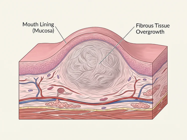
An oral fibroma is a benign (non-cancerous) lump of fibrous tissue that forms on the
mouth lining, the soft tissue covering the inside of the cheeks, lips and gums. It
develops at a spot that has been repeatedly irritated and is one of the most common soft tissue
growths in the mouth. Its clinical name is fibrous hyperplasia, meaning a localised overgrowth
of dense fibrous tissue that builds up in response to ongoing irritation.
A fibroma in the mouth is also referred to as an irritation fibroma or a traumatic fibroma.
These are the same condition under different names, each pointing to what caused it rather than
to a separate diagnosis.
Oral Fibroma Symptoms: What It Looks and Feels Like
An oral fibroma typically appears as a smooth, dome-shaped lump that is firm to the touch and
painless. Most are small, commonly under a centimetre across, and they tend to grow slowly
before levelling off rather than continuing to enlarge indefinitely.
Four features tend to stand out:
- Colour – Usually the same pink as the surrounding lining. Some look paler or whitish on the surface, which happens when repeated friction thickens the outer layer.
- Texture – Smooth and rounded, with a firm, rubbery feel rather than a soft or fluid-filled one.
- Base – Either sitting flat against the lining or attached by a narrow stalk, which makes it move slightly when touched.
- Sensation – Painless in most cases. Discomfort usually means the lump is being bitten or caught, not that anything has changed inside it.
Most people discover a fibroma with their tongue before they see it, since these growths sit along the line where the teeth meet the cheek or tongue.
What Causes an Oral Fibroma?
Repeated irritation to one spot on the mouth lining causes an oral fibroma. Something has to physically rub or catch the same patch of tissue over weeks or months for the growth to develop. It is not caused by diet or infection, and there is no established link to oral hygiene.
Common sources of that irritation include:
- Cheek and lip biting — Accidental or habitual biting that keeps catching the same patch of tissue.
- Teeth grinding and clenching — Bruxism can press the cheek and tongue against the edges of the teeth repeatedly, often during sleep. A mouth guard separates the teeth and reduces that contact.
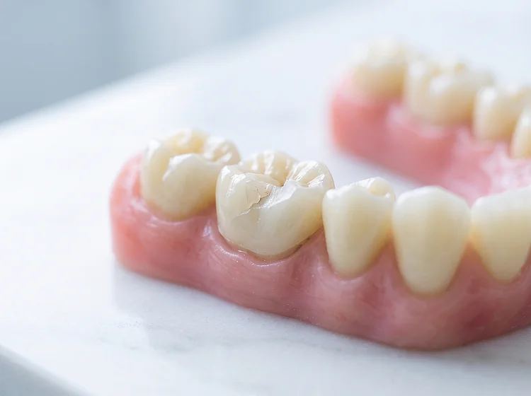
- Ill-fitting dentures or appliances — A denture edge, retainer or aligner tray pressing on one area over months.
- Braces and aligner attachments — Brackets and wires rubbing against the inner cheek or lip, or the small bonded attachments used with clear aligners such as Invisalign.
- A rough or broken tooth — A chipped edge or an old filling with a sharp margin catching the tissue each time the mouth closes.
A single injury heals and closes over within days. An injury that keeps repeating never reaches that point, so the tissue continues repairing itself and fibrous tissue builds up instead.
Where Oral Fibromas Develop in the Mouth
Oral fibromas form wherever the mouth lining meets a source of friction, which is why most appear along the bite line where the upper and lower teeth close together.
Inner Cheek
The inner cheek is where oral fibromas form most often. The lining here sits directly between the upper and lower teeth, so it takes the most friction from chewing and accidental biting. A fibroma inside the cheek often sits level with the back teeth and becomes noticeable once it starts getting caught during meals.
Sides of the Tongue
Tongue fibromas usually appear along the sides rather than on the top surface, since the edges are what rest against the teeth. A fibroma on the tongue tends to be noticed earlier than one elsewhere, because the tongue moves constantly and the lump is felt with almost every word and swallow.
Gums
A fibroma on the gums sits on the tissue between or beside the teeth, usually towards the front of the
mouth. Growths in this position need closer assessment, as two other gum growths, peripheral ossifying
fibroma and pyogenic granuloma, can look similar but are managed
differently.
Gum disease also produces swellings
with a similar appearance, so gum lumps that bleed easily or have grown quickly are worth having
examined sooner rather than later.
Inner Lip
A fibroma on the lip most often forms on the inner surface of the lower lip, which is bitten more often than the upper lip. Growths here are usually visible when the lip is pulled down, so they are typically spotted earlier than fibromas further back in the mouth.
WhatsApp Us Now
Types of Oral Fibroma
Oral fibromas fall into a few distinct types, and the differences matter because they affect how the growth is removed and how likely it is to come back.
| Type | What It Is | Where It Usually Appears |
|---|---|---|
| Irritation fibroma (traumatic fibroma) | The common form, caused by repeated friction or biting | Inner cheek, sides of the tongue, inner lip |
| Giant cell fibroma | A less common form whose trigger is not well established, identified by the distinctive cells seen under a microscope | Gums, tongue, buccal mucosa (inner cheek) |
| Peripheral ossifying fibroma | A gum-specific growth thought to arise from the periodontal ligament that anchors the tooth root, containing hard tissue as well as fibrous tissue | Gums only, usually between the front teeth |
How an Oral Fibroma Is Diagnosed
An oral fibroma is typically diagnosed by examining the lump directly and identifying
the irritation that produced it. This involves checking the size, firmness, colour
and how the growth attaches to the lining, then looking at the teeth, fillings and any appliances
nearby to work out what has been rubbing against the tissue.
An X-ray is usually taken only when the growth sits on the gum, or when the examination points
to involvement of the bone beneath, since neither can be assessed from the surface.
The diagnosis is confirmed after the growth is removed. Appearance alone cannot separate a
fibroma from the other conditions that resemble it, so the tissue is sent for examination under a microscope,
and that analysis establishes what the lump was.
How Is an Oral Fibroma Removed?
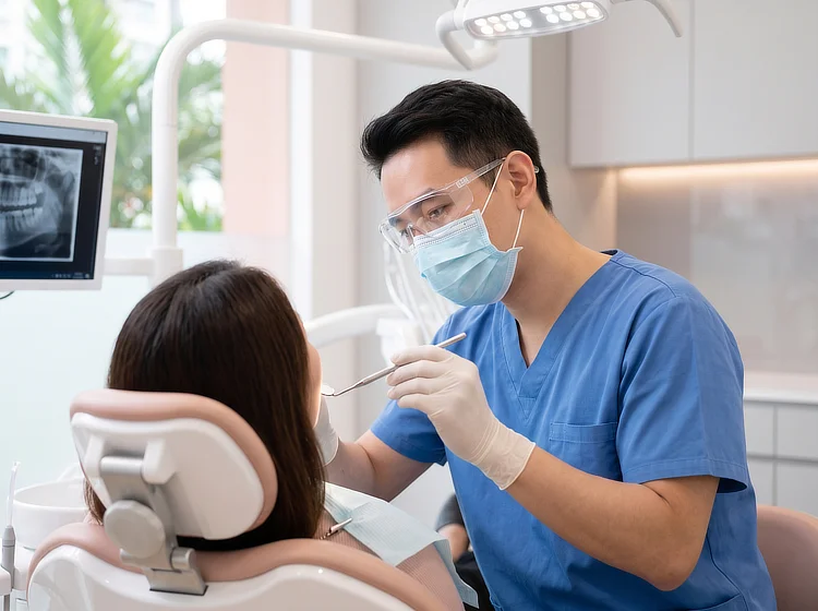
Oral fibroma removal is a minor surgical procedure carried out under local anaesthetic in a single appointment. The whole growth is taken out rather than trimmed, because tissue left behind at the base is what allows a fibroma to reform.
- Numbing the area — Local anaesthetic is given around the growth. You stay awake throughout and may feel pressure or movement, but not pain.
- Removing the growth — The fibroma is taken out in full, including its base, along with a small margin of surrounding tissue so that nothing is left behind.
- Closing the site — Stitches are placed where needed. Smaller sites are sometimes left to heal on their own.
- Laboratory analysis — The removed tissue is sent for examination under a microscope.
Gum growths are removed down to the underlying bone, since any tissue left at the base is what allows
them to return.
Removing the growth is only half the treatment, since the irritation that caused it also needs to be
addressed. That may mean smoothing a sharp tooth edge, adjusting a denture, or recommending a night
guard where grinding is the source.
Recovery After Oral Fibroma Removal
Most people return to normal activity the same day, with the site healing over one to two weeks. Mild
soreness for a few days is usual once the anaesthetic wears off, and it is generally managed with
over-the-counter pain relief.
During healing, the following can help:
- Softer foods — Chew on the opposite side for the first few days while the area is tender.
- Gentle cleaning — Brush around the site rather than avoiding it, since plaque building up there slows healing.
- Salt water rinses — Warm salt water after meals, starting the day after the procedure if advised.
- Leaving the site alone — Probing it with your tongue or fingers disturbs the tissue as it heals.
Stitches either dissolve on their own or are removed at a short follow-up appointment, usually within one to two weeks.
Meet Our Oral and Maxillofacial Surgeon

Dr Albert Lee Ming Hsien
M.D.S. (Oral Maxillofacial Surgery) (Singapore)
F.A.M. (Singapore)
Oral Maxillofacial Surgeon
Dr Albert Lee is an Oral and Maxillofacial Surgery Specialist at True Dental Studio with over 13
years of clinical experience. He holds a Bachelor of Dental Surgery and a Master of Dental
Surgery in Oral and Maxillofacial Surgery from the National University of Singapore, and
received full specialist accreditation in 2018.
Dr Lee previously served as a Consultant at the National Dental Centre Singapore (NDCS), where
he headed the Craniofacial and Oral Implantology Programme and was Surgical Chief at the Oral
and Maxillofacial Clinic in Changi General Hospital. He is a Clinical Assistant Professor
appointed by Duke-NUS.
His clinical interests include dental implants, wisdom teeth surgery, and mandibular
reconstruction, and he has published and presented at local and international conferences.
Most patients come in worried about what the lump might be. In the case of an oral fibroma, one appointment is usually enough to remove the lump and establish what it was. Most people carry on with their day afterwards, with only mild soreness while the site settles.

Dr Albert Lee
Cost of Oral Fibroma Removal in Singapore
At True Dental Studio, the cost of oral fibroma removal depends on the size, site and complexity of the growth, as assessed by our oral and maxillofacial surgeon.
| Treatment | Price* |
|---|---|
| Consultation | $27.25 to $65.40 |
| Second Opinion Consultation | $27.25 to $65.40 |
| Orthopantomogram (OPG) X-ray | $109 |
| Oral Fibroma Removal | Please enquire |
*Prices are NETT and inclusive of GST.
Surgical removal of an oral growth may be claimable through MediSave in
some cases. Contact us to find out more about eligibility or for further information.
When to See a Dentist About a Lump in Your Mouth
Any lump in the mouth that has been present for more than
two weeks is worth having examined. Most turn out to be harmless, and an assessment tells you
what the lump is instead of leaving it unexplained.
Arrange an appointment sooner if you notice:
- A sore that will not heal — An ulcer or raw patch still present after two weeks.
- A hard or fixed lump — One that feels firm to press and does not move against the tissue beneath it.
- Rapid growth — A lump that has enlarged noticeably over days or weeks.
- Unexplained bleeding — Bleeding from the lump without any knock or injury to explain it.
- Colour changes nearby — Red or white patches on the surrounding lining.
- Numbness or persistent pain — Either of these, or a lump in the neck alongside the one in the mouth.
Book a Consultation for Oral Fibroma
Book Oral Fibroma Consultation
Book NowEnquire More Through Email
Email Us NowEnquire More Through WhatsApp
WhatsApp Us NowWhy Choose True Dental Studio?
Friendly Dentist and Team
We treat every patient to the best of our ability. This is why families across Singapore have continued their dental care with us for years.
Minimal Discomfort
We are committed to keeping our patients as comfortable as possible, minimising any discomfort during treatment.
Affordable Treatments
We believe in pricing quality treatments at a competitive rate. We are transparent and upfront with our treatment costs.
Our Smile Scorecard
Frequently Asked Questions
Oral fibromas rarely grow back when the whole growth, including its base, is taken out and the source of irritation is dealt with at the same time. A fibroma that returns usually means the original cause is still there, such as a sharp tooth edge or continued cheek biting. Growths on the gum recur more often, so a lump reappearing at the same spot is worth having checked by your dentist or oral surgeon.
Oral fibromas do not usually bleed on their own, as they are made of dense fibrous tissue rather than blood vessels. Bleeding normally follows an injury, such as biting the lump or catching it while brushing. If a growth bleeds without any knock to explain it, having it examined is advisable, as unexplained bleeding is more typical of other growths.
Infection of an oral fibroma itself is uncommon. The surface can become sore or ulcerated when the lump is bitten repeatedly, and it is that broken surface which may then become infected. Increasing pain, swelling or discharge around the area is worth having checked by a dentist rather than waiting for it to settle on its own.
An oral fibroma is not contagious. It forms from your own tissue responding to repeated irritation, so it cannot be passed on through kissing, sharing food or sharing utensils. No virus or bacterium is involved in how it develops, though a fibroma is only confirmed as one once the growth has been removed and examined under a microscope.
Oral fibromas grow slowly, often over many months, and most settle at a small size, commonly under a centimetre across. Larger ones do occur where the irritation has continued for years. A lump that enlarges noticeably over days or weeks does not fit this pattern, and prompt assessment is advisable in that situation.
Oral fibroma removal is carried out by a dentist or an oral and maxillofacial surgeon, depending on the size and site of the growth. At True Dental Studio, assessment and removal are handled by our oral and maxillofacial surgeon. Larger growths, and those on the gum where the bone beneath may be involved, are the ones most likely to need surgical input.
Visible scarring after oral fibroma removal is unlikely. The soft tissue inside the mouth heals more readily than skin and generally leaves little trace once the site has settled. Some people notice a slightly firmer patch of tissue for a few weeks, which tends to soften as healing finishes.
Oral fibromas do not go away on their own. The lump is established fibrous tissue rather than swelling or inflammation, so there is nothing for the body to reabsorb over time. Removing the source of irritation stops it enlarging further, but does not clear what has already formed. Surgical removal is the only way to clear a fibroma.
Oral fibromas are removed through a minor surgical procedure under local anaesthetic, usually in a single appointment. The whole growth is taken out, including its base, as tissue left behind allows it to reform. The irritation that caused it has to be addressed as well, whether that means smoothing a sharp tooth edge or adjusting a denture.
Oral fibromas are among the most frequently seen soft tissue growths in the mouth. They occur across all age groups and affect adults more often than children, simply because irritation has had longer to accumulate. Their frequency reflects how easily the mouth lining is caught between the teeth during everyday chewing and speaking.
An oral fibroma looks like a smooth, dome-shaped lump, usually the same pink as the surrounding tissue, though some appear paler or whitish where friction has thickened the surface. Most measure under a centimetre across and feel firm rather than soft. Several other conditions look similar, and a fibroma is only confirmed once the growth has been removed and examined under a microscope.
Recovery after oral fibroma removal usually takes one to two weeks for the site to heal, with most people back to normal activity the same day. Mild soreness for a few days is expected once the anaesthetic wears off, and this is generally managed with over-the-counter pain relief. Any stitches placed either dissolve on their own or are removed at a short follow-up appointment within the same period.
A fibroma on the lip is removed the same way as one elsewhere in the mouth, through a minor excision (surgical removal of the growth) under local anaesthetic. The growth is taken out in full, including its base, and the site is closed with stitches where needed. Lip fibromas are usually easy to reach, and the tissue inside the lip generally heals with little visible trace.
No. Attempting to remove an oral fibroma yourself is strictly not advisable. Cutting, tying off or burning the lump at home risks bleeding, infection and scarring, and no rinse or supplement will shrink established fibrous tissue. Self-removal also loses the opportunity to have the tissue examined under a microscope, which is what confirms whether the lump was harmless.
Oral fibromas do not become cancerous. They are made of ordinary fibrous tissue with no tendency to change into anything malignant. A fibroma can only be identified for certain once the growth has been removed and examined under a microscope, since appearance alone cannot separate it from other conditions. A lump present beyond two weeks is worth having looked at.